
PearlBone™ for Clinical Use
An osteoconductive, biomaterial scaffold derived from nacre and engineered to support bone regeneration.

Clinical Applications
-
PearlBone™ is being developed for use in orthopaedic procedures to fill bone voids or defects and support new bone formation. It’s composition and bone-like microarchitecture are designed to facilitate osteoconduction and vascularisation within load-sharing environments, subject to clinical indication and regulatory approval.
-
In traumatic bone injury, PearlBone™ is being developed for use as a bone void filler to support bone regeneration. The material provides a porous scaffold designed to facilitate vascularisation and osteoconduction, and is gradually resorbed and replaced with newly formed bone during the healing process.
-
PearlBone™ is being developed for use in the management of contained bone defects arising from injury, disease or surgical intervention. Its porous microarchitecture is designed to support vascularisation and osteoconduction, facilitating bone ingrowth as the material gradually resorbs and is replaced with native bone over time.

Material Advantages

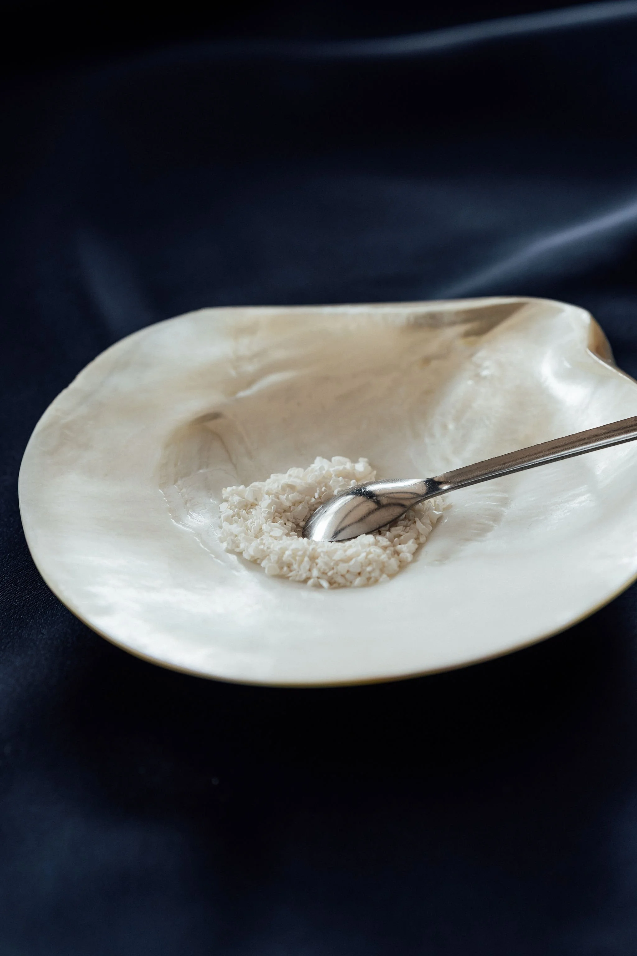
Nacre (mother of pearl) derived from the pearl oyster provides a natural source of calcium carbonate. When combined with hydroxyapatite, it forms a biocompatible material with a bone-like porous microarchitecture designed to support vascularisation and osteoconduction, facilitating new bone formation and progressive replacement with native bone during healing.
Calcium phosphate closely resembles the mineral composition of native bone. In combination with nacre-derived calcium carbonate, it forms a biocompatible material with a bone-like composition, designed to support osteoconduction and facilitate new bone formation.
Research & Validation
PearlBone™ has undergone extensive preclinical evaluation to assess biocompatibility and interaction with host bone tissue. Research to date has focused on material characterisation, scaffold behaviour in biological environments, and early indicators of bone regeneration.
Ongoing studies are progressing through defined preclinical milestones in preparation for future clinical evaluation, subject to regulatory approval pathways.
How PearlBone™ Integrates With Host Bone
PearlBone™ is a natural bone substitute that acts as a biocompatible and osteo-integrating scaffold. When placed within a bone defect, the material provides a supportive structure that allows new bone tissue to integrate within the product.
The porous structure of PearlBone™ is conducive to cellular integration, allowing bone-forming cells to migrate into the scaffold as healing progresses.
Over time, the material is gradually resorbed by the body and replaced with newly formed bone, supporting the natural bone regeneration process.
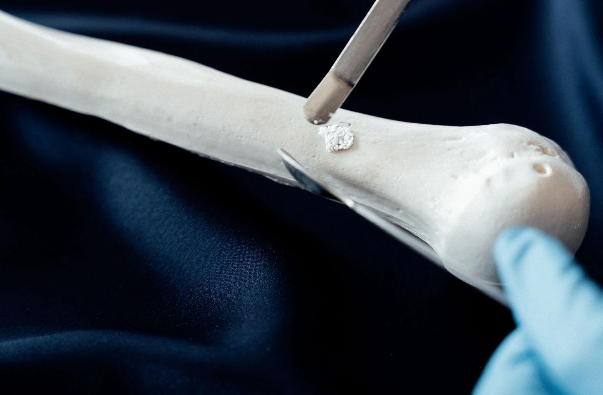
PearlBone™ is placed at the site of bone loss
Bone begins to integrate with the PearlBone™ structure
PearlBone™ is gradually resorbed and replaced by new bone
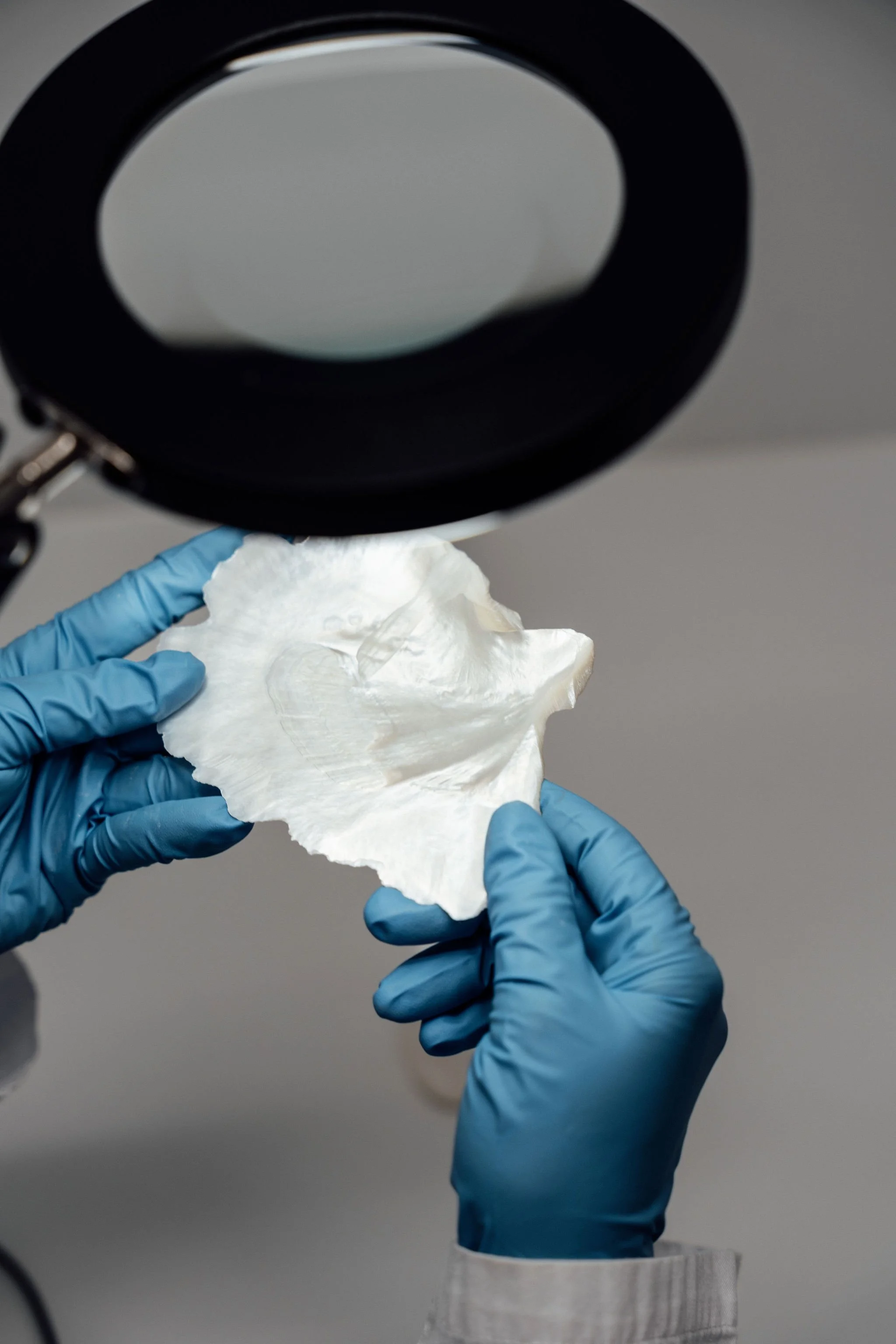
How PearlBone™ Is Studied
Before being used in clinical settings, PearlBone™ is carefully evaluated through laboratory and pre-clinical studies to understand how it behaves and interacts with bone.
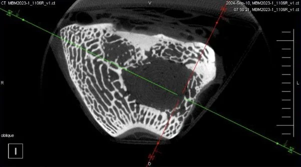
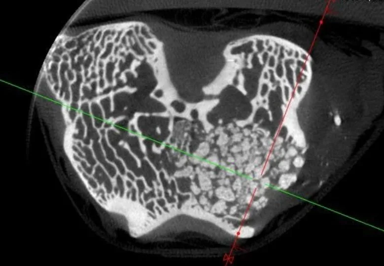
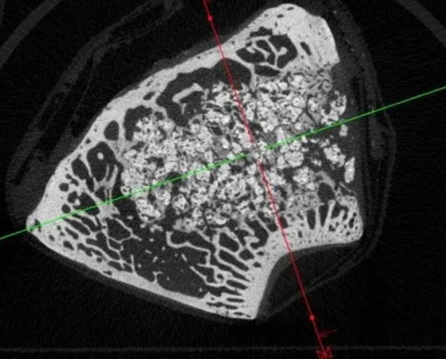
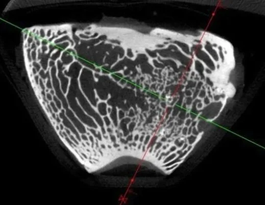
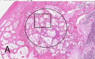
Progressive Bone Regeneration Over Time
Histological analysis demonstrating the integration and replacement of PearlBone™ with bone over 12 weeks.
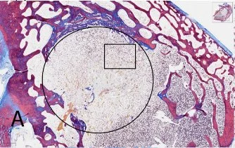
Empty defect.
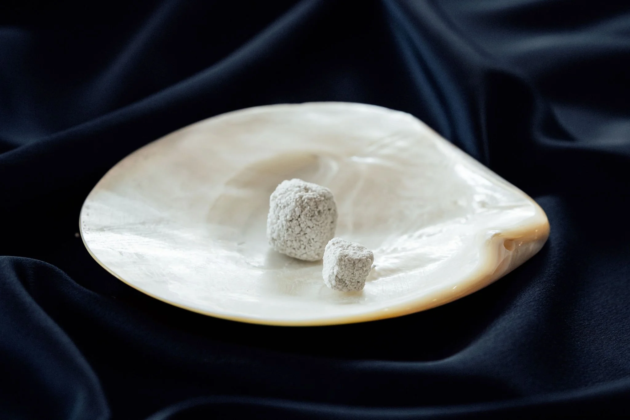
Early implant of PearlBone™ with clearly defined granules.
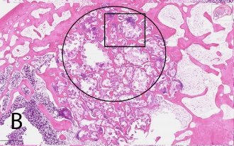
Resorption of PearlBone™ and preliminary bone formation.
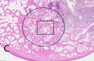
Substantial bone formation and resorption of PearlBone™.
Quality & Safety
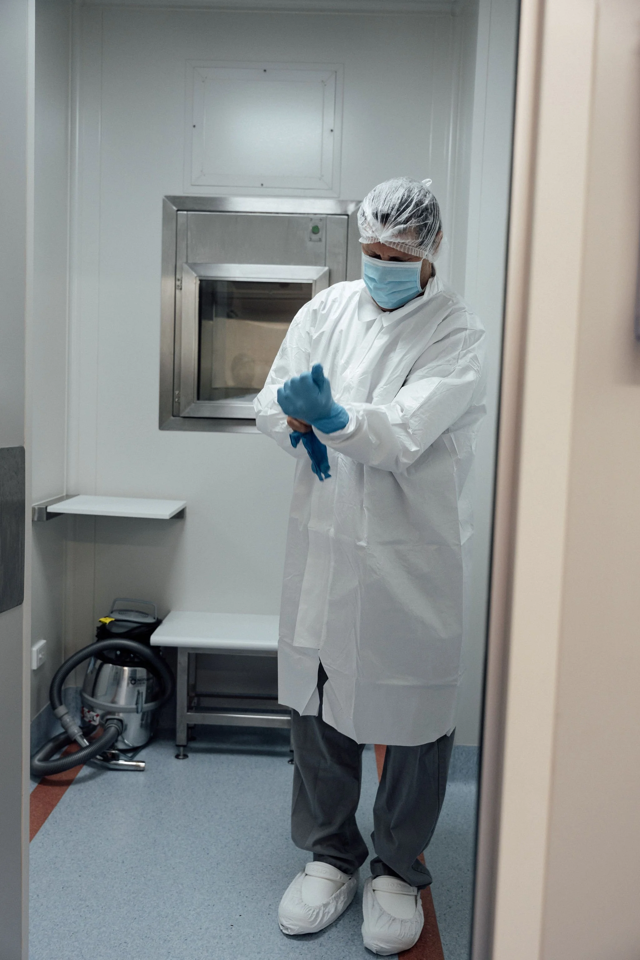
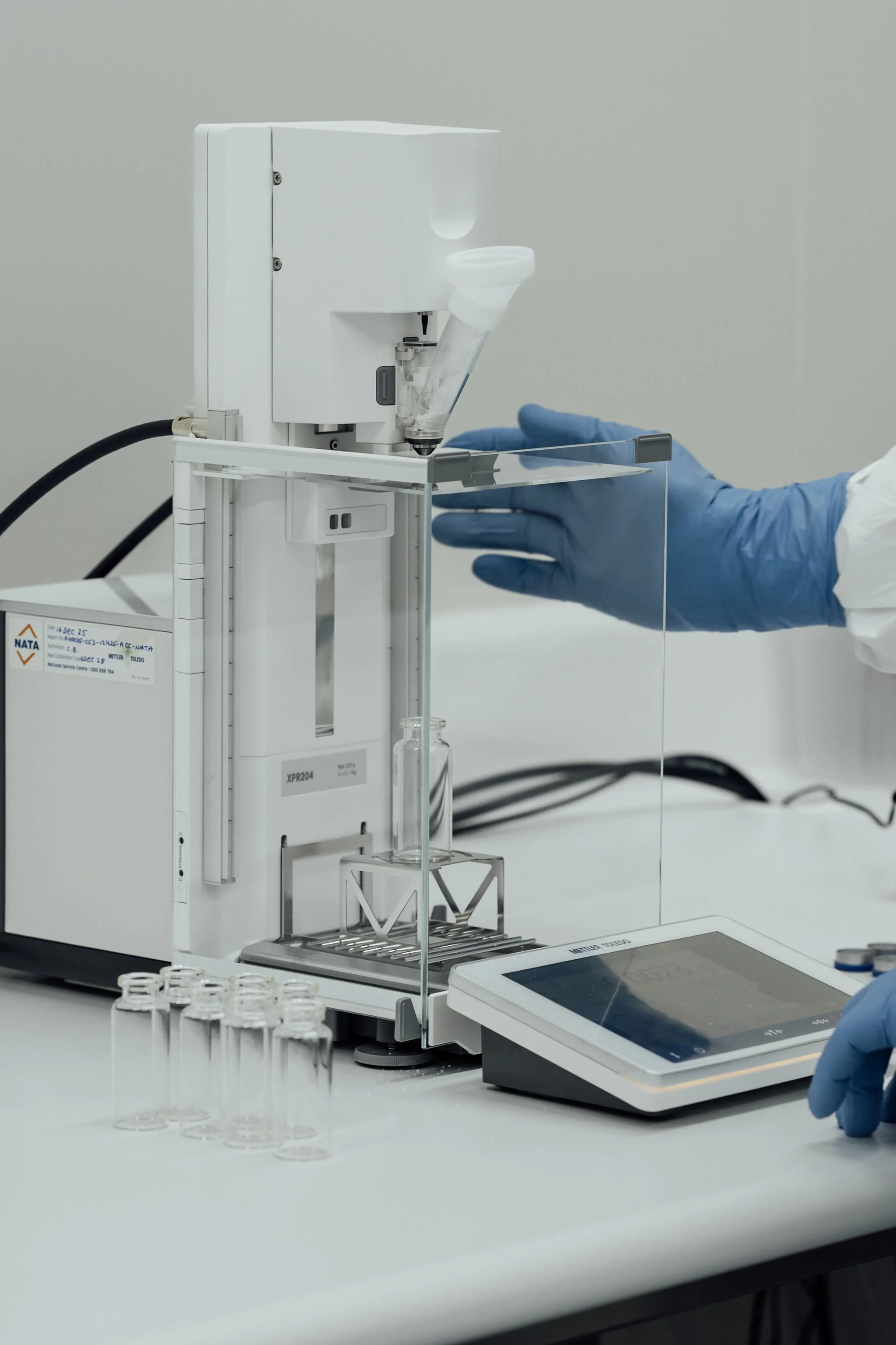
PearlBone™ is manufactured under controlled medical-grade processes designed to meet international quality and safety standards.
GMP-aligned cleanroom manufacturing environment
Quality management system aligned with ISO 13485:2016
Development aligned with FDA and TGA regulatory pathways
Controlled sourcing and traceability of raw nacre material
Validated processing and sterilisation methods
All development activities prioritise patient safety, material consistency, and regulatory compliance.